Abstract
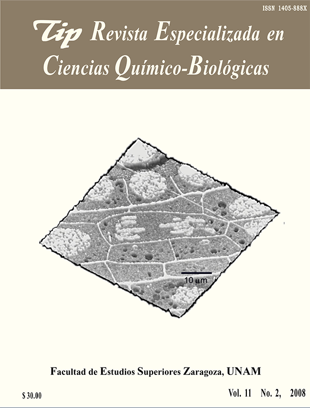
In eukaryotes, cell division generally takes place by mitosis. In previous studies we documented the possibility of studying in situ cell structure by atomic force microscopy, and specially the interphase cell nucleus. Here we show that different stages of mitosis can be visualized with this instrument, therefore offering the possibility to study this phenomenon at the nanoscale.TIP Magazine Specialized in Chemical-Biological Sciences, distributed under Creative Commons License: Attribution + Noncommercial + NoDerivatives 4.0 International.


